1. The Problem with Single-Mode Duct Inspection
Conventional duct inspection is a single-mode activity. The inspector inserts a white-light borescope, advances it through the duct, records what is visible under white illumination, and withdraws the probe. If the findings suggest that UV or thermal investigation is warranted, a separate instrument must be deployed — a separate insertion, a separate pass, and in many cases a separate mobilisation with different equipment.
This sequential approach has two fundamental problems. The first is efficiency: each additional inspection mode requires a separate probe insertion, which multiplies the time on site, the access requirements, and the disruption to the building occupants and HVAC system. For a duct system with multiple access points, a three-mode investigation using separate instruments can take three times as long as a single-mode inspection.
The second problem is evidence quality. When UV and thermal inspections are conducted as separate passes — potentially on different days, under different environmental conditions, with different probe positions — the correlation between findings from different modes is weakened. An inspector who identifies a UV fluorescence anomaly in a second pass cannot be certain that the probe was at exactly the same location as the white-light anomaly documented in the first pass. The spatial correlation that makes multi-spectrum evidence compelling is compromised by the separation of the inspection passes.
SpectraSwitch™ solves both problems by integrating all four imaging modes into a single probe that can switch modes in the field, at the point of interest, without withdrawal. The inspector advances through the duct once, and at any point of interest can switch between white-light, UV 365 nm, UV 405 nm, and thermal/IR imaging — documenting each mode's findings at the same probe position, in the same inspection pass, under the same environmental conditions.
2. What SpectraSwitch™ Is — and How It Works
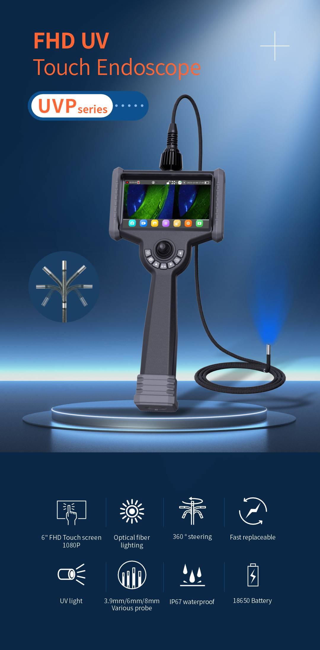
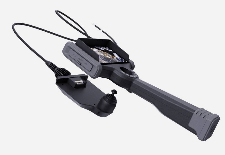
SpectraSwitch™ is the proprietary illumination and imaging switching system integrated into the VD-FID Forensic Inspection Ductoscope. At its core, it is an electronically controlled multi-source illumination system combined with a multi-mode imaging sensor, all housed within the probe tip at the same diameter and form factor as a standard single-mode endoscope.
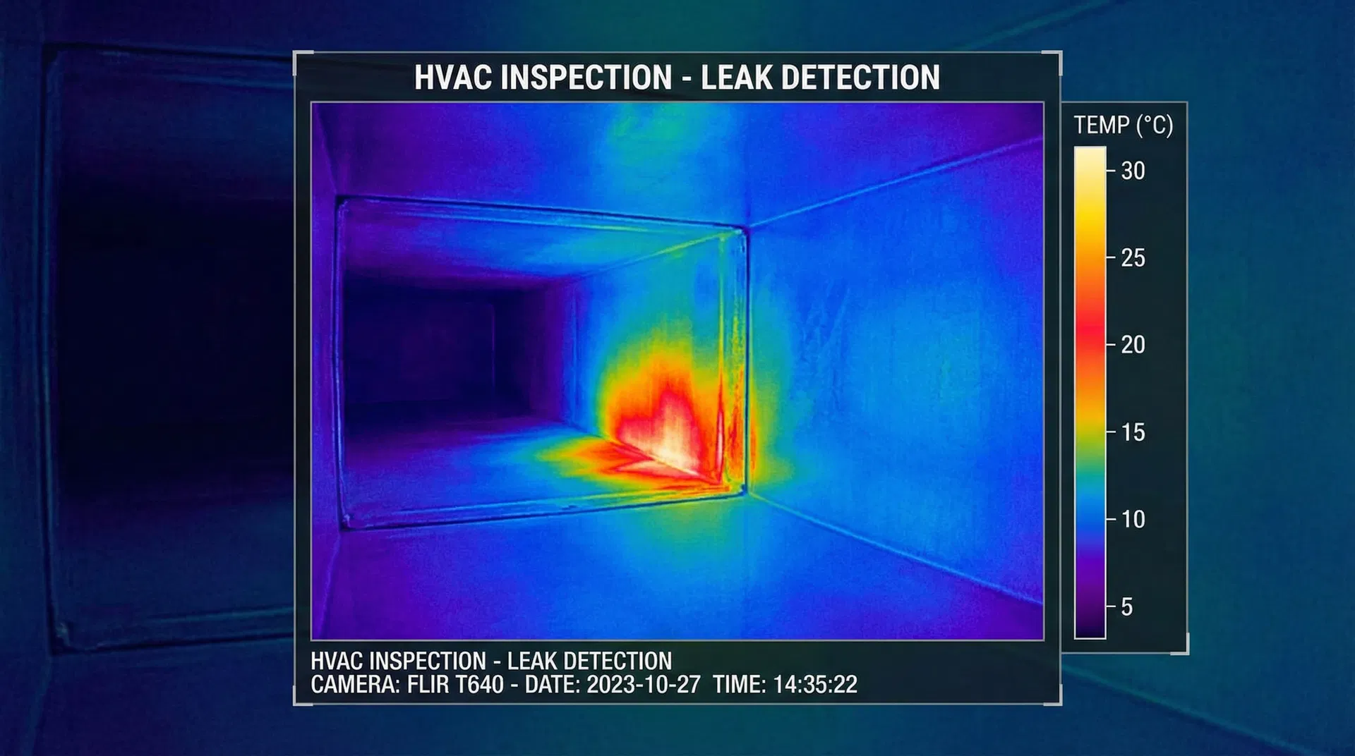
The probe tip contains multiple illumination sources: a high-output white LED array for visible-spectrum imaging, a 365 nm UV LED for near-UV fluorescence excitation, a 405 nm violet-UV LED for broader-spectrum fluorescence characterisation, and an infrared thermal sensor for surface temperature mapping. The controller unit — held by the inspector — provides a mode selector that switches between these sources and configures the imaging pipeline accordingly.
Mode switching is instantaneous. There is no mechanical movement, no lens change, and no delay between modes. The inspector presses a button on the controller, and the probe transitions from white-light to UV 365 nm to UV 405 nm to thermal in under a second. All four modes share the same optical axis and the same field of view, ensuring that images from different modes are spatially registered — a critical requirement for evidence correlation.
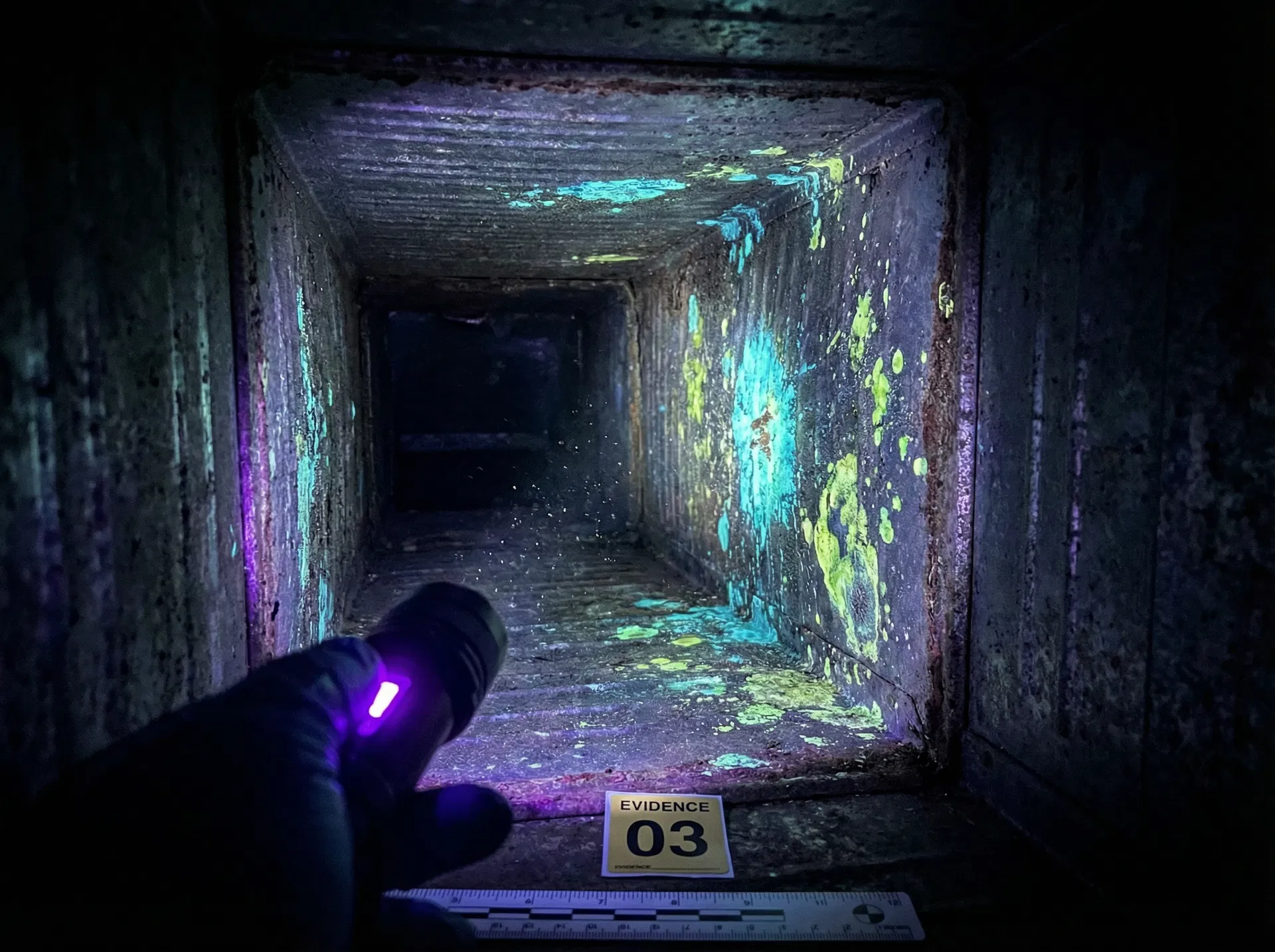
3. The Four Imaging Modes Explained
4. The Integrated Inspection Workflow
The SpectraSwitch™ system is designed around a specific inspection workflow that maximises the value of multi-spectrum evidence while minimising time on site. The workflow proceeds in four stages, each building on the findings of the previous stage.
5. Why Mode-Switching Without Probe Removal Matters
The ability to switch imaging modes without withdrawing the probe is not merely a convenience feature — it is a fundamental requirement for high-quality forensic evidence. When an inspector withdraws a probe and reinserts it to conduct a second-mode inspection, the probe cannot be guaranteed to return to exactly the same position. Duct geometry, probe stiffness, and the inspector's technique all introduce positional uncertainty. In a 20-metre duct run, a second insertion may be off by several centimetres at any given point — enough to miss a localised anomaly or to fail to correlate findings between modes.
For forensic applications, this positional uncertainty is a significant evidentiary weakness. A report that states "UV fluorescence was observed at approximately 12 metres from the access point" and "a thermal anomaly was observed at approximately 12 metres from the access point" — based on two separate probe insertions — cannot definitively establish that these two findings are at the same location. A report based on SpectraSwitch™ in-situ mode switching can state that both findings were documented at the same probe position in the same inspection pass, with the same probe depth reading, under the same environmental conditions. This is a materially stronger evidentiary statement.
The time efficiency benefit is equally significant. A four-mode inspection using SpectraSwitch™ takes approximately the same time as a single-mode inspection — the additional modes add seconds per point of interest, not hours per inspection. A four-mode investigation using separate instruments would require four separate probe insertions, four separate passes, and potentially four separate mobilisations if different instruments are held by different contractors. The SpectraSwitch™ approach reduces a multi-day, multi-contractor investigation to a single inspector, single visit, single pass.
6. Evidence Correlation: When Modes Work Together
The diagnostic power of SpectraSwitch™ is greatest when findings from multiple modes are correlated at the same location. The combination of modes produces a richer evidence picture than any single mode can provide, and the correlations between modes are themselves diagnostically informative.
| White LED | Thermal | UV 365 nm | Interpretation |
|---|---|---|---|
| Normal | Cold spot | No fluorescence | Active leak, no contamination yet — remediate immediately |
| Normal | Cold spot | Strong fluorescence | Active leak with established biofilm — active contamination source |
| Staining | Normal | Strong fluorescence | Past moisture event, active biofilm — residual contamination |
| Staining | Normal | No fluorescence | Past moisture event, dried — historical record only |
| Visible mold | Cold spot | Strong fluorescence | Active mold growth with ongoing moisture — critical finding |
| Normal | Normal | Weak fluorescence | Low-level biofilm, no active moisture — monitor |
This correlation matrix illustrates how the combination of modes produces diagnostic conclusions that no single mode can reach. A thermal anomaly alone does not confirm contamination. UV fluorescence alone does not confirm active moisture. The combination of both — at the same location, in the same pass — provides the evidence for a definitive conclusion about the current state of the duct and the appropriate remediation response.
7. SpectraSwitch™ vs. Separate Instrument Approaches
| Factor | Separate Instruments | SpectraSwitch™ (VD-FID) |
|---|---|---|
| Probe insertions per inspection | 1 per mode (3–4 total) | 1 total |
| Time on site | Multiplied by mode count | Single-pass duration |
| Positional accuracy between modes | Uncertain (re-insertion error) | Exact (same probe position) |
| Evidence correlation quality | Approximate | Spatially co-registered |
| Equipment required | Multiple instruments | Single VD-FID unit |
| Contractor coordination | May require multiple specialists | Single inspector |
| Admissibility for litigation | Weaker (positional uncertainty) | Stronger (co-registered evidence) |
| Post-remediation verification | Separate re-inspection | Integrated in same pass |
8. Applications by Industry and Use Case
SpectraSwitch™ technology is applicable across a wide range of industries and inspection contexts, but its value is particularly pronounced in situations where multi-spectrum evidence is required for decision-making, compliance, or legal purposes.
Infection control investigations require documentation of both the presence of contamination (UV) and the moisture conditions that support it (thermal). SpectraSwitch™ provides both in a single inspection, supporting accreditation compliance and infection control protocols.
Water damage claims involving HVAC systems require evidence of both the water intrusion event (thermal) and any resulting contamination (UV). Co-registered multi-spectrum evidence from a single inspection pass is significantly more compelling than evidence from separate inspections.
Building defect and IAQ litigation requires court-admissible evidence. The spatial co-registration of SpectraSwitch™ findings — documented in a single pass with consistent probe depth readings — provides a stronger evidentiary foundation than evidence from multiple separate inspections.
After duct cleaning and decontamination, SpectraSwitch™ enables a single-pass verification inspection that confirms both the absence of residual contamination (UV) and the resolution of moisture conditions (thermal) — providing a complete clearance record.
9. Technical Specifications and Platform Details
The VD-FID is built on the Videtex VD-UVP/VD-P platforms, which are purpose-designed for multi-spectrum NDT endoscopy applications. The platform integrates the SpectraSwitch™ illumination and imaging system with a high-resolution CMOS sensor, a flexible probe system available in diameters from 0.85 mm to 8 mm and lengths from 0.5 m to 30 m, and a 4.3-inch IPS LCD controller with on-device recording to Micro SD.
| Specification | VD-FID |
|---|---|
| Platform | Videtex VD-UVP / VD-P |
| Imaging modes | White LED, UV 365 nm, UV 405 nm, Thermal/IR |
| UV wavelengths | 365 nm + 405 nm |
| Thermal resolution | < 0.5°C |
| Probe diameter | 0.85 mm – 8 mm |
| Probe length | 0.5 m – 30 m |
| Image sensor | HD CMOS |
| Display | 4.3" IPS LCD |
| Video output | 1080p HD |
| Storage | Micro SD (up to 128 GB) |
| Battery life | 4+ hours continuous |
| IP rating | IP67 probe tip |
SpectraSwitch™ Included as Standard
- Four imaging modes in one probe
- Instantaneous mode switching (< 1 second)
- Spatially co-registered multi-spectrum evidence
- Probe diameters 0.85–8 mm, reach to 30 m
- Court-admissible documentation capability